
FACS ANALYSIS LICENSE
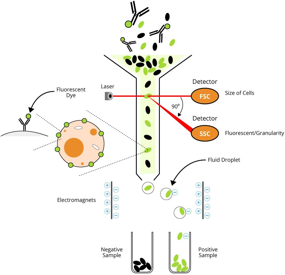
Technical support is also provided for analyses of flow and imaging cytometry data for publication, presentation, and inclusion in grant applications, management of cytometric data (storage, archiving, and retrieval), and management of a site license for low-cost post-acquisition analysis software. Facility personnel aid investigators in creating efficient and cost-effective experimental designs, through optimizing cytometry-specific reagent and fluorochrome selection, and offer assistance in operation of analysis instruments. The Facility is capable of cell sorting (sterile, at speeds up to 30,000 cells/sec) from homogeneous or mixed cell populations based on up to 48 fluorochromes, sorting up to six separate populations simultaneously, including human-derived samples at BSL-2 level.
FACS ANALYSIS PROFESSIONAL
To learn more about getting your flow cytometry data published and to get access to all of our advanced materials including 20 training videos, presentations, workbooks, and private group membership, get on the Flow Cytometry Mastery Class wait list.The Flow Cytometry Shared Resource provides investigators with the technological resources and professional assistance for high quality, multiparameter flow cytometry analyses and sorting.

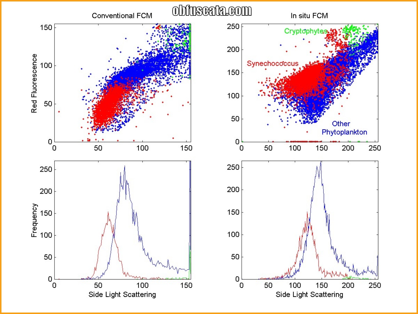
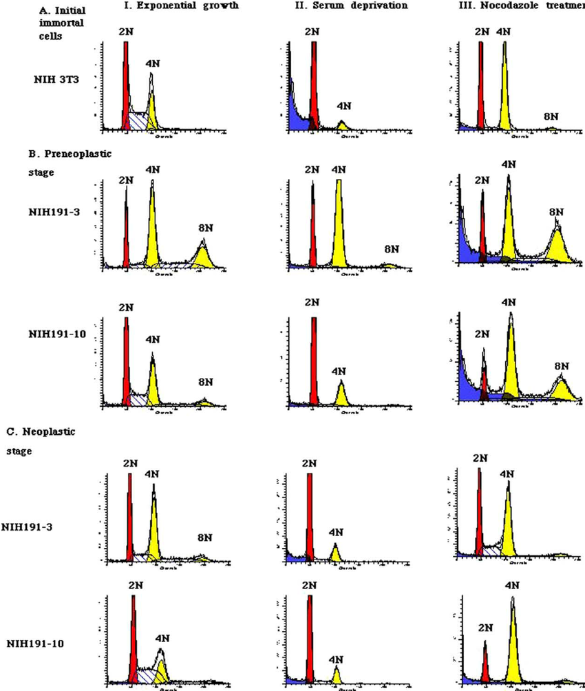
While it provides pretty pictures and colorful layouts, the meat of the data are the numbers ― percentages of populations, fluorescent intensity levels and the like ― these are what will convince the reader that the hypothesis tested is valid and well thought-out. The flow cytometry data that forms the basis of the conclusions should be presented clearly and concisely. When preparing figures for publication, the scientific question and hypothesis that forms the basis of the paper must be central and all the figures must be in support of that. The above presentation of the gating strategy is valuable for dispelling that myth that gating is a subjective art form.Īs new automated analytical techniques become more widespread, they will also help in addressing this issue while adding a level of confidence that the data extracted for downstream statistical analysis has come from a robust, vetted process. Typically, figures are shown with data from different conditions shown on one graph, often with an offset as below… These plots show the intensity of expression versus the number of events. Histograms tend to be the most abused of figures for presenting flow cytometry data. Here are the benefits and drawbacks of popular flow figures to consider when presenting your data: 1. Their purpose is to extract numeric values (such as percent positive or median fluorescent intensity) from the data ― the real value of the data to be presented. The huge caveat with falling in love with any of these types of plots is in knowing the plots used for flow analysis are more often than not a means to an end.

dot plots) and even higher order plots (3D plots, SPADE trees, etc.). Presenting the data in the best possible format, highlighting your results while avoiding glitz that can make the integrity of your data suspicious, is key.Īt first glance, flow cytometry data is very visual.Īnalysis techniques rely on presentations using univariate (a.k.a. There is so much data and so little time that it is essential to present information in the clearest, most concise way.Īs Einstein once said: “Everything must be made as simple as possible. Today, the expectation is that you’ll choose the right flow figures from all that are available, selecting the ones that reflect your data accurately and without confusion. Good handwriting (formerly known as proper penmanship) and drawings might have been enough to convince peers in the distant past, but not today. The presentation of your data must be clear.Īs such, choosing the right flow figures to communicate your data is essential. The goal of any scientific process, as you know, requires the communication of the data that supports or refutes the hypothesis under testing.īefore it is deemed worthy of publication, it must survive the process of peer review ― where the data is laid bare before a group of experts in the field who judge the material impartially (usually) and in secret ― then pass judgment on the suitability of the information for publication. “It would be possible to describe everything scientifically, but it would make no sense it would be without meaning, as if you described a Beethoven symphony as a variation of wave pressure.”


 0 kommentar(er)
